What is an X-ray image and why is it like a shadow?
An X-ray image is a two-dimensional projection. A generator produces X-ray photons, they pass through the dog's body, and a detector on the far side records how many arrive. Tissue stops photons in proportion to its density and atomic composition: bone absorbs strongly, fat and fluid less, air almost not at all. What reaches the detector is therefore a map of attenuation, and the image is read as light and dark rather than as a picture of structures in their true positions.
The shadow comparison is accurate in one important respect. Just as a hand held in front of a lamp casts a flat outline with no depth information, a radiograph collapses everything along the beam path onto one plane. A rib and a lung lesion behind it overlap in the same pixels. Radiographers manage this by taking at least two views at right angles, which lets the reader reconstruct position mentally. The comparison is also used in patient-facing explanations of how an X-ray image, shadow is formed, because the same principle applies whether the subject is a person or an animal.
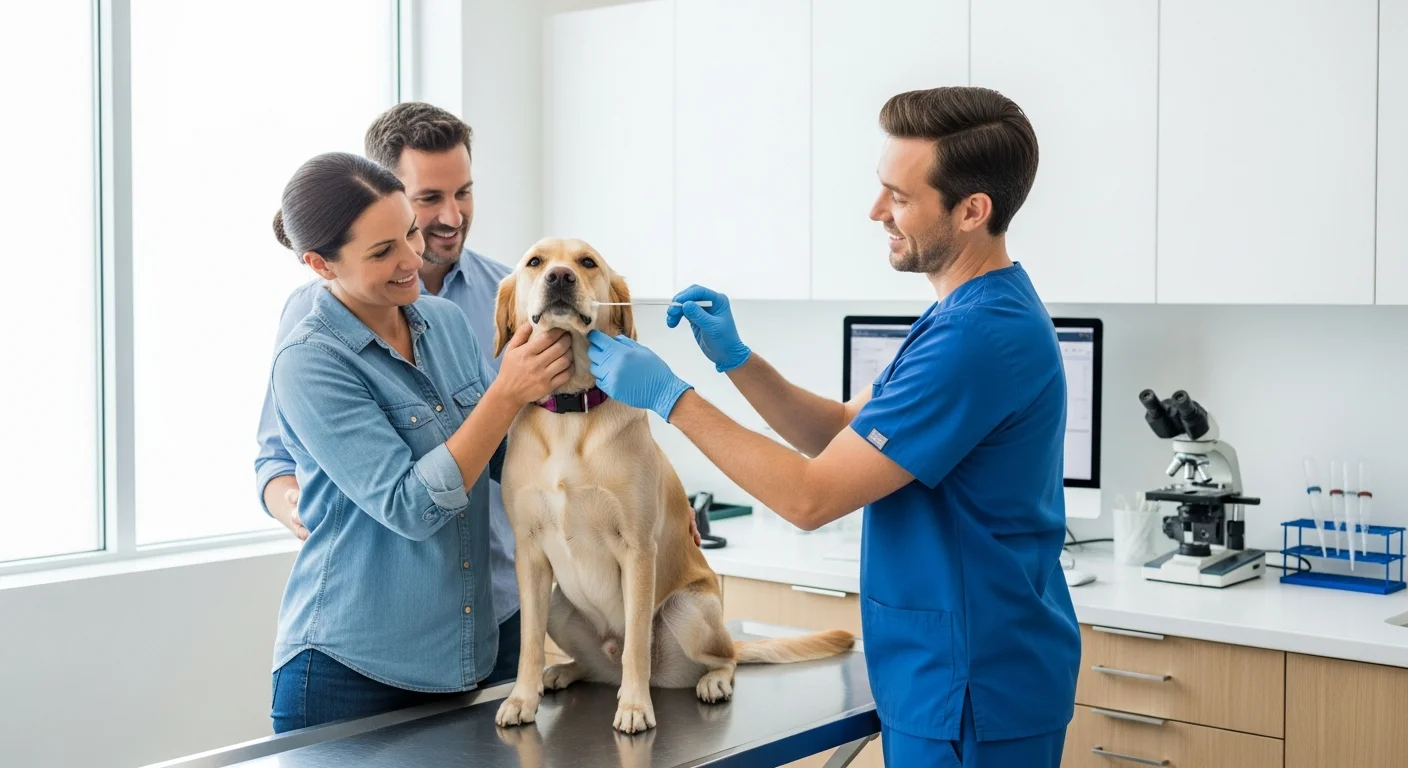
For a dog, positioning matters as much as technique. A lateral view of the thorax and a ventrodorsal view are standard, and the dog must be still, often under light sedation, because motion blurs the projection. Radiography is fast, widely available and inexpensive relative to cross-sectional methods, which is why it is usually the first step for a cough, a lameness or a suspected foreign body.
How does a CT scanner create a computed slice?
A CT scanner also uses X-rays, but it does not produce a single shadow. The tube and detectors rotate around the dog while the table moves through the gantry, collecting attenuation measurements from hundreds of angles. A computer then reconstructs these into cross-sectional images, the slices. The word computed refers to that reconstruction step: the slice is calculated from the projection data, not photographed directly.
Each slice is a thin slab, typically a few millimetres, and the reader can scroll through the stack or reformat it into other planes. Because the data are three-dimensional, a CT study separates structures that a radiograph would superimpose. This is why CT is chosen for nasal disease, complex fractures, spinal detail, pulmonary nodules and surgical planning. The trade-off is radiation dose, which is higher than for a single radiograph, and the need for general anaesthesia or heavy sedation so the dog remains motionless for the duration of the scan.
Contrast agents are often given intravenously during CT. They alter attenuation in vessels and organs and can show a mass's blood supply or a leak. The imaging service checks renal function and history before administering contrast, and the owner is told beforehand that a cannula will be placed and that the injection may feel warm.
Why does an MRI scan not use ionising radiation?
MRI does not use X-rays at all. It places the dog in a strong static magnetic field, which aligns the magnetic moments of hydrogen nuclei in tissue water. A radiofrequency pulse briefly disturbs that alignment, and as the nuclei relax they emit a signal that receiver coils detect. The images are built from the timing and strength of those signals, which differ between tissues. Because the energy involved is radiofrequency rather than ionising, MRI does not carry the radiation risk associated with radiography and CT.
The safety profile is different, not absent. The strong magnetic field means ferromagnetic objects must never enter the scan room, and implants, pacemakers and some metallic foreign bodies must be screened before the appointment. Anaesthesia is required, and the scan takes longer than CT, often thirty to sixty minutes for a full study. MRI gives the best soft-tissue contrast of the three methods, which is why it is used for brain, spinal cord, joint and soft-tissue questions where CT would be less informative.
What does the referral letter ask for?
The referral is a written clinical question, not a general request for pictures. A useful letter states the presenting signs and their duration, the relevant history and previous treatments, the specific region to be imaged, and the clinical question the referring vet wants answered. It also lists current medication, any known reaction to contrast, and relevant blood results.
The imaging service uses this to decide the protocol: which modality, which views or sequences, whether contrast is needed, and how much time to book. A vague request such as scan the abdomen produces a less targeted study than a question such as is there a pancreatic mass, and where. In the United Kingdom, the imaging service also applies justification and optimisation duties under the Ionising Radiation (Medical Exposure) Regulations 2017, meaning each exposure must be individually justified and kept as low as reasonably practicable while still answering the question.
What is the owner told before and after the appointment?
Before the appointment, the owner is normally told where to go, what time to arrive, whether food should be withheld, which medications to give or skip, and whether the dog will be sedated or anaesthetised. For contrast studies, the owner may be asked about previous reactions and about kidney disease. For MRI, the owner is asked about implants, previous surgery and any metal the dog may have swallowed.
On the day, the dog is admitted, examined, and usually has an intravenous catheter placed. The owner leaves and is contacted when the dog is recovering. After the study, the owner is given a discharge summary covering what was done, how the dog tolerated it, and when to expect the report. The formal report goes to the referring vet, who interprets it in the context of the whole case and discusses the findings with the owner. Images are stored and can be shared for a second opinion.
How the methods compare in practice
The three methods answer different questions. Radiography is quick, cheap and good for bone, gas, fluid lines and gross organ size. CT adds cross-sectional detail and three-dimensional reconstruction at the cost of a higher dose. MRI avoids ionising radiation and gives the best soft-tissue contrast, but is slower, less widely available and more expensive.
For a dog owner, the practical differences are timing, cost, anaesthesia and what the result can show. The referral letter determines which of these is appropriate, and the report returns to the vet who knows the dog. Understanding how each image is built makes the sequence of decisions easier to follow, from the first radiograph to a scan that answers a narrower question.
Imaging answers a structural question, but it does not describe how a dog moves between appointments or how comfortable that movement is. Owners often ask what happens after a diagnosis, when the clinical picture is clear but the dog still guards a limb or tires on stairs. Rehabilitation sits in that gap, and it is a separate field with its own practitioners, costs and limits. A short overview of hands-on work and movement explains what canine massage, TTouch, physiotherapy and underwater treadmills involve, who practises them in Germany, and what a session costs.
Further Reading